Understanding Hoof Anatomy: A Complete Guide for Livestock Owners
You don’t need to be a farrier to understand hoof anatomy. If you own livestock—horses, cows, goats, or sheep—knowing how a healthy hoof should look and function makes daily care easier. Hooves tell stories. The way they grow, wear, and react to movement shows what’s happening inside. Learning the basics of animal foot anatomy and hoof structure means fewer surprises, better communication with your trimmer, and animals that stay sound longer.
Why Knowing Hoof Anatomy Is Important
A healthy-looking animal can still have hidden issues. And by the time your livestock starts limping, damage may already be done. But if you know what normal hooves feel like, small changes won’t slip past you.
Even minor shifts in the sole and frog can signal deeper trouble. Understanding the parts of the hoof helps you spot problems early. It also gives you more confidence in daily cleaning and trimming. You’re not just guessing—you’re observing and connecting details. That awareness comes from paying attention to hoof anatomy every day.
When you know the layout of internal vs external hoof parts, talks with farriers and vets get easier. You can describe issues clearly—like heat near the coronary band or a softened frog. That kind of accuracy helps everyone act faster.
Over time, checking for flare or uneven wear becomes second nature. Those habits lead to consistent care that keeps hooves healthier and animals more comfortable.
Spotting Subtle Signs Early
Knowing where and how hoof problems begin makes checks more effective. Early signs include cracks climbing up the hoof wall, bad smell in the frog’s groove, or faster wear on one side. They seem small at first but often lead to lameness. Familiarity with hoof anatomy allows you to catch these issues while they’re still easy to manage.
Making Daily Care Smarter
Hoof cleaning changes once you understand each part’s role. Instead of rushing, you start noticing frog texture, sole hardness, and edges of the hoof wall. It only takes seconds but adds up over time. Each detail connects back to hoof anatomy and the way each structure supports the animal.
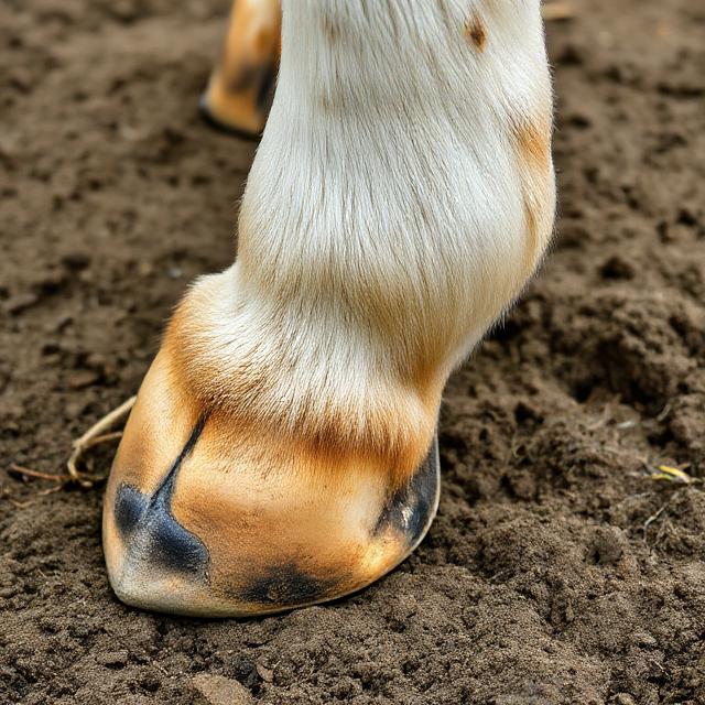
External Parts of the Hoof Explained
The outside of the hoof does more than look tough—it supports weight, handles shock, and protects what’s inside.
- Hoof wall: the strong outer barrier that shields internal tissues.
- Sole: the curved base that helps carry weight and defend the inner foot.
- Frog: the triangle-shaped shock absorber that assists circulation.
- Heel bulbs: soft, rounded areas behind the frog that add cushion.
- Bars: ridges that give extra support and keep the hoof from collapsing.
- White line: the narrow band where the sole meets the wall; it often shows early signs of imbalance.
Each part helps with balance, grip, and movement. When something changes—like a stretched white line or contracted heel—it disrupts how the whole hoof functions. Watching these parts over time helps prevent future problems. Knowing the outer hoof anatomy helps you recognize damage before it worsens.
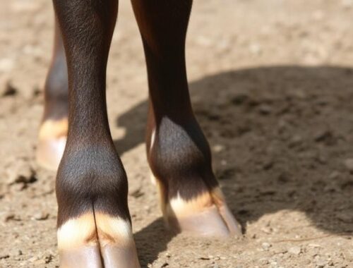
Internal Structures of the Hoof
Inside the hoof capsule lies a complex system of bone, tissue, and circulation. The hoof capsule surrounds these parts like a strong shell. Every time the animal steps, the internal structures absorb shock and help pump blood. Understanding this level of hoof anatomy helps you connect surface changes with what’s happening inside.
| Structure | Horse | Cow | Goat/Sheep |
|---|---|---|---|
| Hoof Wall | Thick and strong | Thinner than horses | Hard but brittle |
| Frog | Large, supports circulation | Smaller, less defined | Small and narrow |
| Digital Cushion | Deep and firm | Softer, especially in dairy cows | Less developed |
| Sole | Tough and concave | Flatter surface | Prone to overgrowth |
| P3 (Coffin Bone) | Fully enclosed in hoof capsule | Split between claws | Narrow claws with space |
Species share the same parts, but the shape and feel vary. The digital cushion in horses is firmer and thicker than in goats. Goats often grow hoof wall faster and chip more easily.
The Role of the Digital Cushion
Sitting just above the frog, this pad of fat and fiber softens each step. In fit animals, it feels thick and springy. Without enough movement, it shrinks and loses shock absorption. That makes the hoof more vulnerable to injury. Balanced trimming and plenty of exercise help maintain its strength. It’s one of the many internal elements that shows how hoof anatomy supports movement and comfort.
How Anatomy Affects Hoof Function
A hoof isn’t just a shell—it’s a dynamic system. How it’s shaped affects how pressure spreads. If one part isn’t working right, everything else compensates.
- Long toes put stress on tendons and delay breakover.
- Low heels reduce shock absorption and tire joints faster.
- Narrow frogs block expansion and slow circulation.
- Uneven soles shift posture and wear down joints.
- Thin hoof walls fail to guard sensitive inner areas.
Noticing how these parts relate helps you spot early signs of imbalance. It also helps explain changes in gait, like shorter strides or toe-first landings. When hoof anatomy is off, the whole animal feels it.
Daily Care That Respects Hoof Anatomy
Balanced hooves are easier to maintain when you know what balance looks like. A shrinking frog, chipped sole, or widened white line all show stress. Being aware of these signals makes trims and checks more focused and useful. Recognizing early changes is easier when you understand how every detail in hoof anatomy plays a role.
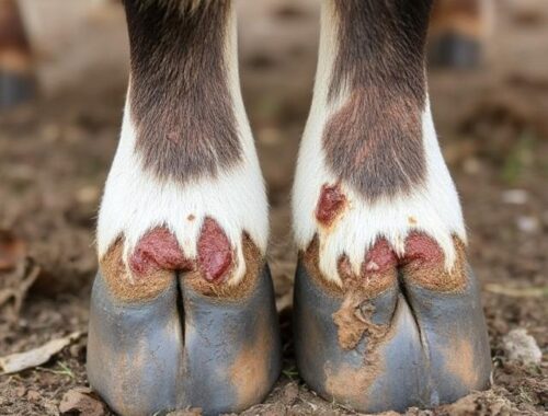
Common Anatomical Weak Points
Certain hoof areas break down faster than others. The white line, for instance, stretches under stress and allows bacteria in. The frog can soften in wet weather and become infected. Heels may contract if movement is limited. Over-trimming thins the sole and exposes tissue. Swelling around the coronary band is often a warning sign.
Once you know where problems usually start, prevention becomes much easier. Knowledge of hoof anatomy makes these patterns more obvious and helps you act sooner.
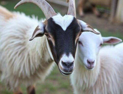
Species Differences: Horse vs Cow vs Goat
Hoof types vary by species. Horses have a single hoof made for motion. Cows walk on two claws that grow unevenly without trimming. Goats and sheep also have two claws, but theirs are smaller and more fragile. That makes them prone to splitting and chipping.
Trimming schedules differ. Goats often need attention every few weeks. Cattle may go longer, but claw balance must be watched. Horses may get flares if they stand too long or don’t move enough.
Understanding differences in hoof anatomy by species makes your care more effective. These structural traits influence everything from how often you trim to how animals move across terrain.
Using Anatomy Knowledge in Daily Hoof Care
Once you know the parts of the hoof and how they work, daily care shifts. You stop looking at hooves as just something to clean. They become indicators. A thin frog might show low movement. A stretched white line could point to early laminitis.
When you understand hoof capsule structure, trimming makes more sense. Knowing how the sole and frog work together helps you judge terrain better. That insight keeps hooves strong and movement steady. Routine care feels less random when it’s grounded in real hoof anatomy.
You don’t need to know every detail of internal vs external hoof parts to help your animals. Just understanding basic hoof anatomy, including the hoof wall, digital cushion, and frog, lets you spot early changes. That awareness turns into better care, healthier hooves, and fewer long-term problems. These small insights make a big difference in your livestock’s comfort and movement every day.


You May Also Like
Common Hoof Diseases in Livestock and How to Treat Them
Comparing Cloven and Single Hooves: Anatomy Across Species